Sheep Brain Labeling Worksheet for Biology Students

Understanding the Sheep Brain: A Labeling Worksheet for Biology Students
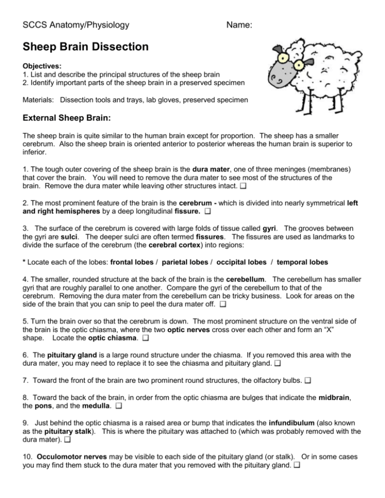
As biology students, understanding the structure and function of the brain is crucial for grasping various biological concepts. One of the most effective ways to learn about brain anatomy is by labeling diagrams of actual brains. In this worksheet, we will focus on the sheep brain, which is commonly used in biology classes due to its similarities to the human brain.
Why Use a Sheep Brain?
Sheep brains are often used in educational settings because they are relatively inexpensive and easy to obtain. Additionally, the sheep brain is similar in structure to the human brain, making it an excellent model for learning about brain anatomy. By labeling a sheep brain diagram, you will gain a deeper understanding of the brain’s structure and function, which will help you in your biology studies.
Sheep Brain Labeling Worksheet
Below is a diagram of a sheep brain. Use the following key to label the different structures:
- Cerebrum
- Cerebellum
- Brainstem
- Frontal lobe
- Parietal lobe
- Temporal lobe
- Occipital lobe
- Hypothalamus
- Thalamus
- Midbrain
- Pons
- Medulla oblongata
- Corpus callosum

 |
Key:
- Cerebrum: The largest part of the brain, responsible for processing sensory information, controlling movement, and managing higher-level brain functions.
- Cerebellum: Located at the base of the brain, the cerebellum coordinates muscle movements and maintains posture and balance.
- Brainstem: Connecting the cerebrum to the spinal cord, the brainstem regulates basic functions such as breathing, heart rate, and blood pressure.
- Frontal lobe: One of the four lobes of the cerebrum, the frontal lobe is responsible for decision-making, problem-solving, and motor control.
- Parietal lobe: Another lobe of the cerebrum, the parietal lobe processes sensory information related to touch and spatial awareness.
- Temporal lobe: The temporal lobe is involved in processing auditory information and is also important for memory and language.
- Occipital lobe: The occipital lobe is primarily responsible for processing visual information.
- Hypothalamus: A small region of the brain that regulates body temperature, hunger, and thirst.
- Thalamus: The thalamus acts as a relay station for sensory information, helping to process and transmit signals to the cerebral cortex.
- Midbrain: Part of the brainstem, the midbrain is involved in auditory and visual processing.
- Pons: Also part of the brainstem, the pons helps regulate sleep and arousal.
- Medulla oblongata: The lowest part of the brainstem, the medulla oblongata controls vital functions such as breathing and heart rate.
- Corpus callosum: A bundle of nerve fibers that connects the two hemispheres of the cerebrum, allowing them to communicate.
Labeling the Diagram
Using the key provided, label the different structures on the sheep brain diagram. Start by identifying the cerebrum, cerebellum, and brainstem, and then work your way through the rest of the structures.
Tips and Notes
- Make sure to label each structure clearly and accurately.
- Use a pencil to label the diagram, as you may need to make corrections.
- If you are unsure about a particular structure, refer to the key or consult with your instructor.
- Take your time and work methodically through the diagram.
💡 Note: Be sure to label the structures on the diagram in the correct locations. If you are unsure, consult with your instructor or refer to a brain anatomy textbook.
As you complete this labeling worksheet, you will gain a deeper understanding of the sheep brain’s structure and function. This knowledge will help you in your biology studies and provide a solid foundation for future learning.
Summing Up
Labeling a sheep brain diagram is an excellent way to learn about brain anatomy and understand the structure and function of the brain. By completing this worksheet, you have demonstrated your knowledge of the sheep brain’s different structures and their roles. Remember to review the key and diagram regularly to reinforce your understanding of brain anatomy.
Why is the sheep brain used in biology classes?
+The sheep brain is used in biology classes because it is relatively inexpensive and easy to obtain. Additionally, the sheep brain is similar in structure to the human brain, making it an excellent model for learning about brain anatomy.
What is the function of the cerebellum?
+The cerebellum coordinates muscle movements and maintains posture and balance.
What is the corpus callosum?
+The corpus callosum is a bundle of nerve fibers that connects the two hemispheres of the cerebrum, allowing them to communicate.


