5 Key Facts About Microscopic Anatomy of Skeletal Muscle

Understanding the Microscopic Anatomy of Skeletal Muscle
Skeletal muscle is one of the three major muscle types in the human body, alongside smooth and cardiac muscle. It plays a crucial role in movement, posture, and overall physical function. At the microscopic level, skeletal muscle has a unique structure that enables it to perform its functions efficiently. Here are five key facts about the microscopic anatomy of skeletal muscle:
1. Muscle Fibers and Fascicles
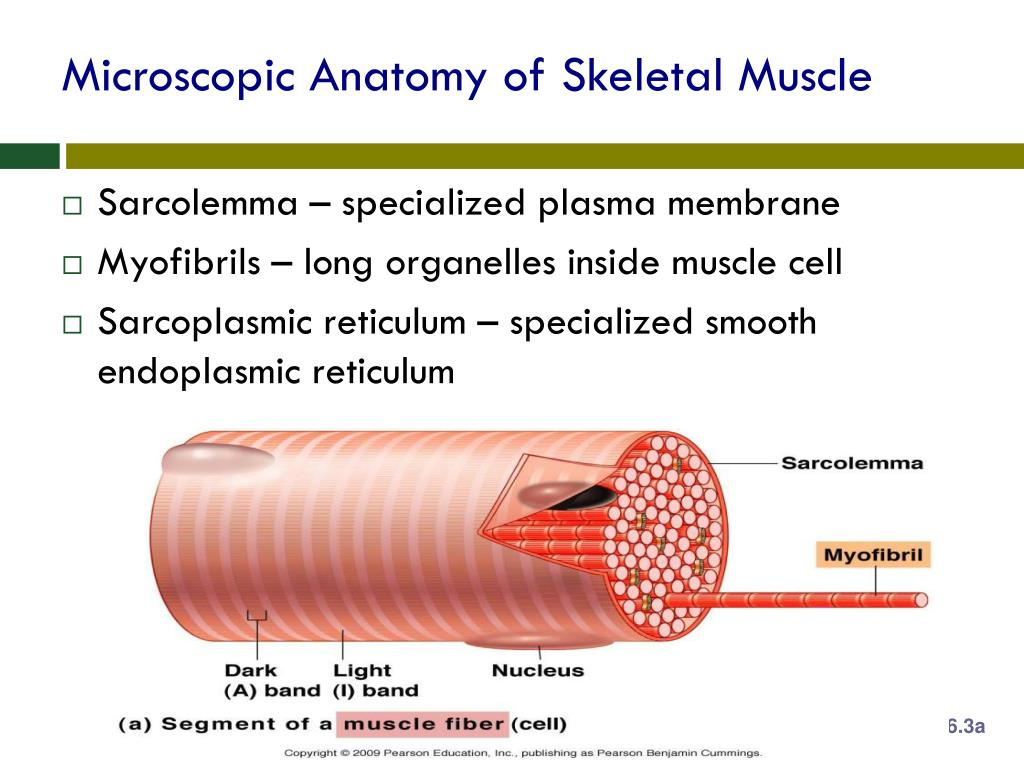
Skeletal muscle is composed of numerous muscle fibers, which are long, multinucleated cells. These fibers are grouped together in bundles called fascicles, surrounded by a connective tissue sheath known as perimysium. Each fascicle contains between 10 to 100 muscle fibers, and the number of fascicles within a muscle can range from a few to several hundred.
2. Sarcolemma and Sarcoplasm
The muscle fiber is surrounded by a thin membrane called the sarcolemma, which regulates the exchange of ions and nutrients between the muscle fiber and the surrounding environment. Inside the sarcolemma lies the sarcoplasm, a fluid-filled cytoplasm that contains various organelles, including mitochondria, ribosomes, and lysosomes. The sarcoplasm also houses the myofibrils, which are the contractile units of the muscle fiber.
3. Myofibrils and Sarcomeres
Myofibrils are the contractile elements of the muscle fiber, composed of repeating units called sarcomeres. A sarcomere is the smallest functional unit of the muscle fiber, measuring approximately 2-3 μm in length. It consists of two types of filaments: thick filaments (myosin) and thin filaments (actin). The arrangement of these filaments within the sarcomere determines the muscle fiber’s contractile properties.
4. T-Tubules and Sarcoplasmic Reticulum
T-tubules (transverse tubules) are narrow, tube-like structures that penetrate the muscle fiber, allowing for the rapid transmission of action potentials. They are closely associated with the sarcoplasmic reticulum, a type of smooth endoplasmic reticulum that stores and releases calcium ions. The T-tubules and sarcoplasmic reticulum work together to regulate muscle contraction by controlling the release of calcium ions.
5. Motor Endplates and Neuromuscular Junctions
Motor endplates are specialized regions on the surface of the muscle fiber that receive signals from motor neurons. They are part of the neuromuscular junction, a synapse between the motor neuron and the muscle fiber. The motor endplate contains acetylcholine receptors, which bind to the neurotransmitter acetylcholine released by the motor neuron. This binding event triggers a series of chemical reactions that ultimately lead to muscle contraction.
🔍 Note: Understanding the microscopic anatomy of skeletal muscle is essential for appreciating the complexities of muscle function and the various factors that influence muscle contraction.
In conclusion, the microscopic anatomy of skeletal muscle is a remarkable and intricate system that enables muscle contraction and movement. By understanding the structure and function of muscle fibers, fascicles, myofibrils, sarcomeres, T-tubules, and motor endplates, we can gain a deeper appreciation for the complexities of muscle biology and the factors that influence muscle function.
What is the main function of the sarcoplasmic reticulum in skeletal muscle?
+The sarcoplasmic reticulum stores and releases calcium ions, which play a crucial role in regulating muscle contraction.
What is the difference between a muscle fiber and a fascicle?
+A muscle fiber is a single, multinucleated cell, while a fascicle is a bundle of muscle fibers surrounded by a connective tissue sheath called perimysium.
What is the function of the T-tubules in skeletal muscle?
+T-tubules allow for the rapid transmission of action potentials, enabling the muscle fiber to contract quickly and efficiently.


